This article covers clinical signs that may be found on the hands during routine clinical examination. The list of clinical signs in this article is by no means exhaustive. Each sign is grouped with the system it is usually associated with (e.g. Janeway lesions and the cardiac system). Other associations are also given.
Notably, it does not cover rheumatologic signs in the hands, such as Heberden’s nodes, or swan neck deformity.
Cardiology
Splinter haemorrhages
Dark red/brown vertical lines seen at the top of the nail. They are small emboli lodged in the nail capillaries damaging vessel walls and causing localised haemorrhage. Causes include trauma, sub-acute bacterial endocarditis, scleroderma, and other autoimmune conditions.

Splinter haemorrhages: Micro-emboli deposited in the nail bed
Osler’s nodes
These painful, small, red lesions appear on the fingers. They are the result of immune complex deposition from bacterial endocarditis.

Osler’s nodes: Patient with bacterial endocarditis 1
Janeway lesions
Painless red spots appearing on the palms. Patients with bacterial endocarditis may develop these lesions from septic emboli causing microhaemorrhage.

Janeway lesions: Patient with endocarditis 2
Fingertip pallor
Fingers appear white and waxy as a result of vasoconstriction or vascular obstruction. Conditions associated with this include peripheral vascular disease, Raynaud’s, Buerger’s disease and CREST syndrome.

Finger pallor: Secondary Raynauds in Sjögrens syndrome 3
Respiratory
Nail clubbing
Nails have increased curvature resulting in the loss of the angle between the nail and nail fold. The exact cause is unclear but patients with clubbing have increased levels of platelets in the microcirculation around the nails resulting in the release of vascular endothelial growth factor (VEGF) and platelet-derived growth factor (PDGF).
Although clubbing can be the result of diseases across multiple systems, it is often related to respiratory pathology when appearing in clinical exams. It is important to remember that clubbing can, in some cases, be a normal finding.
Causes
Respiratory:
- Cystic fibrosis
- Tuberculosis
- Pulmonary fibrosis
- Bronchiectasis
- Bronchial carcinoma
Cardiac:
- Atrial myxoma
- Cyanotic heart disease
- Endocarditis
- Pericarditis
Gastrointestinal:
- Malabsorption
- Inflammatory bowel disease
- Liver cirrhosis

Nail clubbing 4
Carbon-dioxide retention flap (Asterixis)
Asterixis is a coarse tremor best seen with the patient’s wrists extended. It can be caused by hypercapnia, typically in conditions such as chronic obstructive pulmonary disease (COPD) and hypoventilation secondary to reduced consciousness level. Asterixis can also be caused by hyperammonaemia and uraemia.

Asterixis
Hepatology
Tendon/tuberous xanthomata
Yellow nodules typically noted over the dorsal aspects of the hands. Lipid-laden macrophages are deposited as the result of hypercholesteraemia.

Xanthomata over the knee joint 5
Leukonychia
These white, horizontal bands across the nails are the result of low albumin levels in the blood. This can be the result of either decreased albumin synthesis, as seen in liver disease, or the result of increased albumin loss, usually the result of kidney malfunction (nephrotic syndrome). Leukonychia may also be the result of chemotherapy treatment.

Leukonychia
Palmar erythema
Reddening of the palms from vasodilation. Associated pathology includes liver disease and resultant oestrogen excess, and hyperthyroidism.
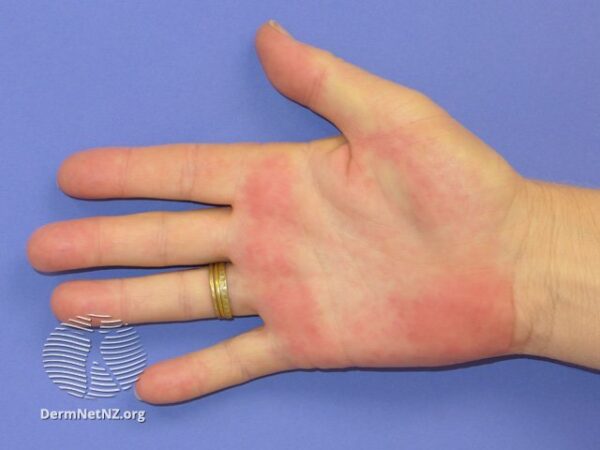
Palmar erythema 6
Asterixis
This flapping-tremor is best elicited by asking the patient to fully extend their wrists and then close their eyes. It can be a sign of hepatic encephalopathy, usually related to long-term alcohol excess, and indicates high levels of ammonia in the blood sufficient to impact on the motor centres of the brain. Other causes include hypercapnia (as discussed previously) and uraemia.
Miscellaneous
Koilonychia
Classically described as ‘spoon-shaped’, these nails appear hollowed out or concave. They are associated with low levels of iron; this could be severe iron-deficiency anaemia, haemochromatosis, fungal infection, acromegaly, hypothyroidism or malnutrition.

Koilonychia 7
Nail pitting
Small indentions of the nail. Pathophysiology is unclear but this pitting is most commonly seen in patients with psoriasis.

Nail pitting in a patient with psoriasis 8
Dupuytren’s contracture
The 4th digit (or ring finger) is in a fixed-flexed position, with the tendon raised and visible on the palmar surface. This appearance is the result of thickening and shortening of the palmar fascia, possibly due to local hypoxia. Associations include smoking, alcohol use, diabetes, manual labour and trauma.

Dupuytren’s contracture 9
References
1. Roberto J. Galindo [GFDL (http://www.gnu.org/copyleft/fdl.html) or CC BY-SA 4.0 (https://creativecommons.org/licenses/by-sa/4.0)], from Wikimedia Commons
2. Warfieldian [CC BY-SA 4.0 (https://creativecommons.org/licenses/by-sa/4.0)], from Wikimedia Commons
3. Intermedichbo [CC BY-SA 3.0 (https://creativecommons.org/licenses/by-sa/3.0) or GFDL (http://www.gnu.org/copyleft/fdl.html)], via Wikimedia Commons
4. Desherinka [GFDL (http://www.gnu.org/copyleft/fdl.html) or CC BY-SA 4.0 (https://creativecommons.org/licenses/by-sa/4.0)], from Wikimedia Commons
5. Min.neel [CC BY-SA 3.0 (https://creativecommons.org/licenses/by-sa/3.0)], from Wikimedia Commons
6. DermNet, Available here: https://www.dermnetnz.org/topics/palmar-erythema/ [Creative Commons Attribution-NonCommercial-NoDerivs 3.0 (New Zealand) at https://creativecommons.org/licenses/by-nc-nd/3.0/nz/legalcode]
7. Corey Heitz MD. Flickr [https://www.flickr.com/photos/coreyheitzmd/15023020192] . Licence [CC2.0 https://creativecommons.org/licenses/by/2.0/].
8. DermNet, Available here: https://www.dermnetnz.org/topics/nail-psoriasis-images/ [Creative Commons Attribution-NonCommercial-NoDerivs 3.0 (New Zealand) at https://creativecommons.org/licenses/by-nc-nd/3.0/nz/legalcode]
9. Frank C. Müller [CC BY-SA 4.0 (https://creativecommons.org/licenses/by-sa/4.0)]
10. Examination of the Hand (The Hand in Diagnosis). Available here: http://stanfordmedicine25.stanford.edu/the25/hand.html#terrys
11. First Aid for USMLE Step 1 (2016 edition)
12. Oxford Handbook of Clinical Medicine
Editor
Fiona Kirkham
The post Clinical Signs of the Hands appeared first on Geeky Medics.