A renal system examination involves looking for clinical clues and signs related to end-stage renal disease (e.g. fistula, dialysis catheter, renal transplant), renal failure complications (e.g. fluid overload, uraemia), transplant immunosuppression side effects (e.g. tremor, striae, steroid facies) and possible cause of renal disease (e.g. diabetes, hypertension, polycystic kidney disease).
This guide provides a generic overview of the potential signs you may identify in a patient with renal disease. The commonest renal patients you’ll come across will be those with polycystic kidney disease, a kidney transplant and/or end-stage renal disease on dialysis.
Check out the renal system examination mark scheme here.
Introduction
- Wash your hands
- Introduce yourself
- Confirm the patient’s name and date of birth
- Explain the procedure – “This will involve me looking at your hands, arms, face and stomach. I’d also like to have a feel and listen to your stomach with a stethoscope. This will require exposing the stomach area.”
- Gain consent and consider a chaperone
- Position the patient (initially supine at 45°)
- Enquire about pain or other concerns prior to beginning the examination
General inspection
Around the bedside
- Oxygen, IV drips, medications, haemodialysis machine, peritoneal dialysis machine
- Urinary catheter, drains (e.g. nephrostomy)
- Fluid chart
- Capillary blood glucose machine – diabetes
Patient
- Consciousness – altered consciousness level may be present in severe or end-stage renal disease
- Nutritional/fluid status – look for evidence of cachexia due to protein-energy wasting (PEW), dehydration or fluid overload
- Overt Cushingoid appearance – may be due to steroids for renal transplant immunosuppression or other conditions (e.g. minimal change glomerulonephritis)
- Uraemic complexion – yellow skin colour may indicate advanced chronic kidney disease
- Breathlessness may be due to fluid overload (e.g. due to anuric state)
- Hyperventilation may be due to metabolic acidaemia secondary to renal failure
Hands
Flapping tremor (asterixis):
- Can indicate uraemia due to acute or end-stage renal disease
- Other causes include CO2 retention and hyperammonemia
Pinprick skin marks from capillary blood glucose readings:
- Suggestive of underlying diabetes
Nail features:
- Beau’s lines – transverse grooves of the nailbed may indicate severe systemic illness
- Muehrke’s lines – transverse white lines of the nail may suggest hypoalbuminaemia
- Lindsay’s half-and-half nail – brown distal fingernails may indicate advanced chronic kidney disease
- Splinter haemorrhages in endocarditis from either valve disease or from dialysis catheter-associated infections
Gouty tophi:
- Common in advanced chronic kidney disease
Skin turgor:
- Useful as part of an overall hydration assessment (decreased in dehydration)
Arms
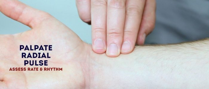
Radial pulse:
- Perform a brief assessment of rate and rhythm
Inspect for excoriation:
- Pruritus can indicate uraemia
Inspect for bruising:
- May be due to excessive corticosteroid use (i.e. immunosuppression) or platelet dysfunction observed in uraemia
Inspect for obvious warts or skin cancers:
- Associated with immunosuppression (i.e. renal transplant patients)
Inspect for an arteriovenous fistula in the wrist (radio-cephalic fistula) and antecubital fossa (brachio-cephalic or brachio-basilic fistula) or the presence of a synthetic PTFE graft in the antecubital fossa (now commonplace in haemodialysis):
- Indicates requirement of haemodialysis
- Look for needle marks indicating that the vascular access is being used
- Palpate for thrill and auscultate for a bruit (both absent if the fistula is thrombosed or surgically ligated such as after renal transplantation)
Offer to measure blood pressure (not on the side of an AV fistula):
- Elevated in hypertension, chronic kidney disease, transplant rejection or as a side effect to steroids, tacrolimus or ciclosporin for renal transplant immunosuppression
- Rarely, pulsus paradoxus (change in BP >10mmHg during breathing) can occur due to uraemic cardiac tamponade (associated with low jugular venous pressure)
Face
General
Skin colour & skin lesions:
- Yellow skin colour (uraemic complexion) – may indicate chronic renal failure
- Lesions associated with immunosuppression – SCC, BCC, in sun-exposed areas or scars from their excision, gingivostomatitis (HSV) around the mouth
Cushingoid facial appearance:
- May be due to steroids for renal transplant immunosuppression or treatment of glomerulonephritis.
Hypertrichosis:
- Can occur in patients on ciclosporin for renal transplant immunosuppression
Hearing aid:
- May suggest Alport’s syndrome
Eyes
Conjunctival pallor:
- Anaemia due to chronic renal failure
Calcification of cornea (band keratopathy):
- Rarely due to elevated blood phosphorus in renal failure
Periorbital oedema:
- Can be seen in nephrotic syndrome
Mouth
Gingival hypertrophy:
- A potential side-effect of immunosuppressants for renal transplant
Uraemic fetor (breath smelling like ammonia):
- Occurs due to uraemia and is seen in end-stage renal disease
Neck
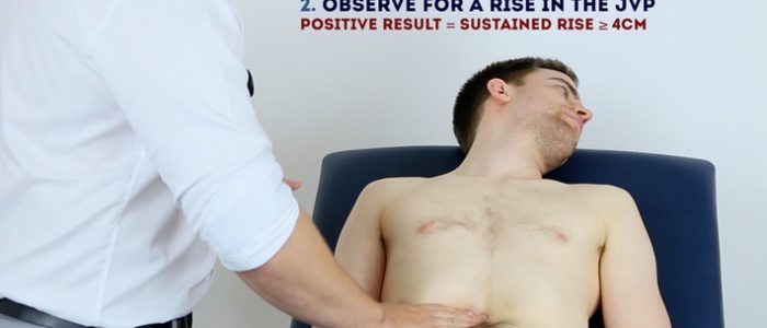
Assess jugular venous pressure (JVP)
1. Ensure the patient is positioned at 45°
2. Ask the patient to turn their head away from you
3. Observe the neck for the JVP – located inline with the sternocleidomastoid
4. Measure the JVP – number of centimetres from the sternal angle to the upper border of pulsation
A raised JVP may indicate – fluid overload, right ventricular failure, tricuspid regurgitation
Other
Look for the presence of an indwelling dialysis catheter at the base of the neck or front of the chest wall.
Inspect for a small horizontal scar at the base of the neck:
- It may be a parathyroidectomy scar (performed for renal hyperparathyroidism)
- May have scars around the base of neck from previous dialysis catheters
Chest
Inspection
Obvious warts or skin cancers:
- Can be caused by immunosuppression (i.e. in the context of renal transplant)
Excoriations:
- Pruritus can indicate uraemia
Bruising:
- May be due to steroids for renal transplant immunosuppression
Percuss
Dull area of percussion on the chest wall:
- May occur if hypoalbuminaemia or fluid overload causes pleural effusion
Palpate
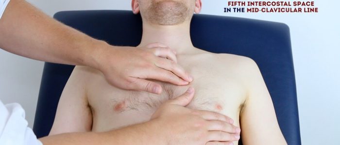
Palpate the apex beat:
- Normally located in the 5th intercostal space in the mid-clavicular line
- Palpate the apex beat with your fingers (placed horizontally across the chest)
- Displacement may indicate fluid overload or heart failure
Auscultate
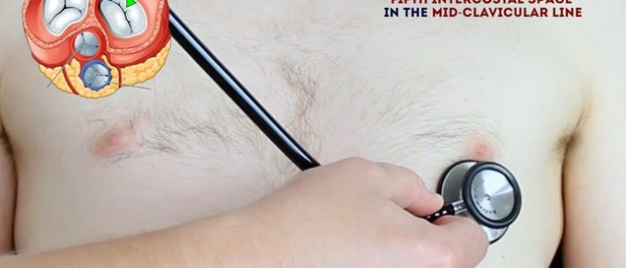
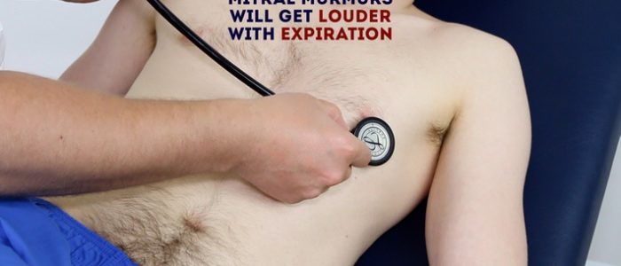
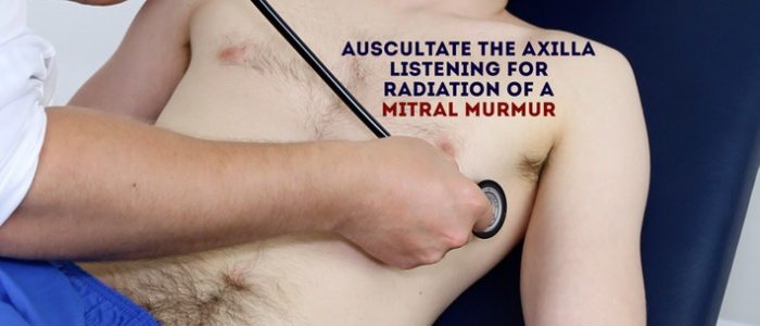
Auscultate heart sounds:
- Note any added sounds which may indicate heart failure and/or fluid overload
- A friction rub may suggest uraemic pericarditis
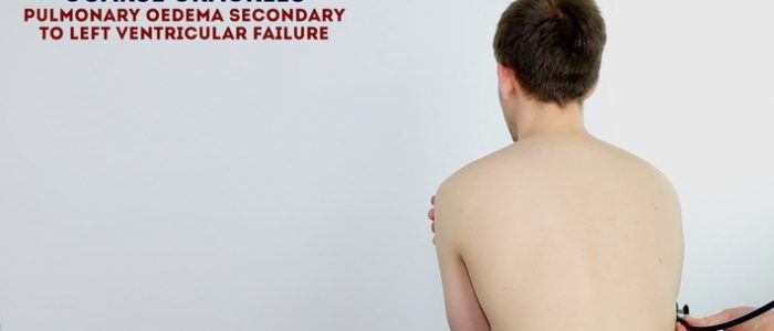
Auscultate lung bases:
- Bibasal crackles may suggest pulmonary oedema secondary to fluid overload
- Vocal resonance may indicate a pleural effusion (e.g. nephrotic syndrome)
Abdomen
Inspection
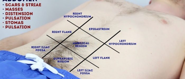
Position the patient flat with their abdomen fully exposed. Look for striae associated with corticosteroid use or abdominal wall oedema associated with fluid overload.
Abdominal distension:
- Intrabdominal masses (i.e. polycystic kidneys)
- Ascites (i.e. nephrotic syndrome)
- Indwelling peritoneal dialysis fluid (look for peritoneal dialysis catheter)
Scars:
- Rutherford-Morrison (‘hockey-stick’) scar for renal transplant
- Bilateral iliac fossae scars from a simultaneous pancreas-kidney transplant (for a patient with type 1 diabetes)
- Peritoneal dialysis catheter insertion scar near the umbilicus
- Nephrectomy scar in the flank
- Lipodystrophy marks from insulin injections for diabetes
Other:
- Tenckhoff catheter in situ for peritoneal dialysis
- Nephrostomy tube(s) in situ
Palpation
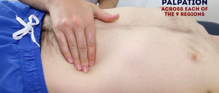
Light and deep abdominal palpation
Palpate the 9 abdominal regions lightly then deeply:
- If a palpable mass is present, consider a polycystic kidney
- A palpable mass and scar in the right or left iliac fossa is suggestive of renal transplant
Ballot the kidneys
1. Place your left hand behind the patient’s back, at the right flank
2. Place your right hand just below the right costal margin in the right flank
3. Press your right hand’s fingers deep into the abdomen
4. At the same time press upwards with your left hand
5. Ask the patient to take a deep breath
6. You may feel the lower pole of the kidney moving inferiorly during inspiration
7. Repeat this process on the opposite side to assess the left kidney
If the kidney is palpable describe the size and consistency:
- Bilaterally enlarged, ballotable kidneys can occur in polycystic kidney disease or amyloidosis
- A unilaterally enlarged, ballotable kidney can be due to a renal tumour
Percussion
Shifting dullness
1. Percuss from the centre of the abdomen to the flank until dullness is noted
2. Keep your finger on the spot at which the percussion note became dull
3. Ask the patient to roll onto the opposite side to which you have detected the dullness
4. Keep the patient on their side for 30 seconds
5. Repeat your percussion in the same spot:
- If fluid was present (ascites) then the area that was previously dull should now be resonant
- If the flank is now resonant, percuss back to the midline, which if ascites is present, will now be dull (i.e. the dullness has shifted)
Auscultation
Auscultate for a renal bruit just above and lateral to umbilicus on both sides:
- A bruit may indicate renal artery stenosis (a possible cause of hypertension or renal failure)
Other
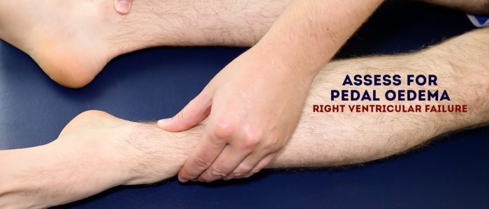
Palpate for sacral and lower limb oedema:
- May indicate fluid overload in nephrotic syndrome or glomerulonephritis or renal impairment
To complete this examination
- Thank the patient
- Allow them time to re-dress
- Wash hands
- Summarise findings
Suggest further assessments and investigations
- Blood pressure – if not already performed (not on the side of the AV fistula)
- Fundoscopy – to assess for the presence of hypertensive or diabetic retinopathy
- Urinalysis – to screen for evidence of infection and to assess for haematuria/proteinuria associated with glomerular disease
- 24-hour urine collection – to assess various urinary compounds and the degree of proteinuria or spot urine protein-creatinine ratio or albumin-creatinine ratio
- MSU – if a urinary tract infection is suspected
- U&Es – to assess renal function
- Bicarbonate – to check for acidaemia
- Bone profile – to assess the levels of calcium, phosphate and PTH (secondary and tertiary hyperparathyroidism)
Reviewed by
Dr Ian Logan
Consultant Nephrologist
Dr Paul Callan
Consultant Cardiologist
The post Renal System Examination – OSCE Guide appeared first on Geeky Medics.

