Quantcast
Channel: OSCE – Geeky Medics
Viewing all 223 articles
Browse latest View live

Peak Expiratory Flow Rate (PEFR) measurement

$
0
0

Peak expiratory flow rate (PEFR) measurement often appears in OSCE’s and involves a combination of both information giving (explaining the procedure) and the practical clinical skill of performing PEFR measurement. The communication skills aspect of this station is where most of the marks lie, so ensure you provide a clear explanation of the procedure to the patient, checking understanding and summarising as you go along.

Introduction

Introduce yourselfstate your name & role

Check patient details - name & DOB

Wash hands


Explain the purpose of the procedure:

“I’d like to assess your breathing”

“This involves measuring how well the air can flow out of the lungs”

“It’s an important test, as it gives an indication of how well your asthma is controlled”


Gain consent - “Do you understand everything I’ve said?” “Are you happy to go ahead with this?”

Measuring PEFR

Ensure you clearly explain & demonstrate each step of the procedure below to the patient:

1. Ensure the PEFR meter is set to zero

2. Sit up straight or stand

3. Take a deep breath (as deep as you can possibly manage)

4. Place your mouth around the mouthpiece of the PEFR meter, ensuring a tight seal with your lips

5. Exhale as forcefully as you possibly can manage

6. Note the PEFR reading

7. Repeat this process  a further 2 times

8. The highest reading of the 3 should be taken as the overall result

To complete the procedure…

Observe patient performing PEFR measurement to ensure good technique

Ask if the patient has any questions regarding PEFR management

Thank patient

Wash hands

The post Peak Expiratory Flow Rate (PEFR) measurement appeared first on Geeky Medics.


Elbow examination – OSCE Guide

$
0
0

Elbow examination can occasionally appear in OSCE’s, so it’s important you’re familiar with it. You should feel confident diagnosing local joint issues such as bursitis, but also be able to identify stigmata of systemic diseases such as psoriasis (plaques) and rheumatoid arthritis (nodules).

Introduction

Wash hands

Introduce yourselfstate your name & role

Confirm patient detailsname & DOB


Explain examination:

“I’d like to examine your elbow”

“This will involve having a look and feel of the joint, in addition to assessing the joints movement”


Gain consent - “Do you understand everything I’ve said?” “Are you happy to go ahead with the exam?”

Gain adequate exposure- ideally you should be able to see the entire limb

Position patientpalms facing forwards with arms by their side (anatomical position)

Look
Front

Scarssuggestive of previous injury / surgery

Swelling / erythema of the joint - may suggest acute injury / inflammatory arthritis / infection

Carrying angle5-15 degrees – females tend to have more significant carrying angles than males

Side

Fixed flexion deformityoften post traumatic 

Olecranon bursitis - the swelling overlying the olecranon is often most noticeable from this angle

Scars / Swelling / Erythema 


Back

Rheumatoid nodulesfirm lumps on the elbow / olecranon – indicate systemic rheumatoid disease

Psoriatic plaqueswell defined pink / red elevated lesions with silvery scale

Feel

Temperaturea particularly warm joint may indicate inflammatory arthritis or infection

Palpate the joint lines –  including the epicondyles & olecranon for any localised tenderness

Move

Assess each of the movements of the elbow joint actively & passively


Elbow flexion - 145º

Elbow extension -  

Pronation – 70º

Supination – 85º


When moving the joint passively assess for crepitus.

Special tests
Medial epicondylitis – a.k.a. “Golfers Elbow”

Ask the patient to actively flex the wrist whilst the elbow is flexed

Localised pain over the medial epicondyle suggests a diagnosis of medial epicondylitis


Lateral epicondylitis – a.k.a. “Tennis Elbow”

Ask the patient to actively extend the wrist whilst the elbow is flexed

Localised pain over the lateral epicondyle suggests a diagnosis of lateral epicondylitis

 To complete the examination…

Thank patient

Wash hands

Say you would…

Examine the joint above & below (shoulder / wrist)

Perform a full neurovascular examination of the upper limbs

Request X-rays of the joint if you suspected pathology

Perform blood tests for markers of inflammation / autoantibodies (e.g. CRP/ Anti-CCP / etc)


The post Elbow examination – OSCE Guide appeared first on Geeky Medics.

Ankle and Foot examination

$
0
0

Ankle and foot examination can occasionally appear in OSCE’s, so it’s important you’re familiar with it. 

Introduction

Wash hands

Introduce yourselfstate your name & role

Confirm patient detailsname & DOB


Explain examination:

“I’d like to examine your ankles and feet”

“This will involve having a look and feel of these joints, in addition to assessing the joints movement”


Gain consent - “Do you understand everything I’ve said?” “Are you happy to go ahead with the exam?”

Gain adequate exposure- both legs should be exposed from the knee down

Position patientask patient to stand up straight with their feet aligned facing forwards

Look
Gait

Is the patient demonstrating a normal Heel strike / Toe off gait?

Is the gait smooth & symmetrical?

Front

Symmetry of feet / ankles

Toe alignment - hallux valgus of the big toe may be noted

Bunions - located at the 1st MTP joint

Toe clawing?

Scarssuggestive of previous injury / surgery

Calluses - may indicate foot / gait deformity or poorly fitting footwear

Swelling / erythema of the foot or ankle - may suggest injury / inflammatory arthritis / infection

Examine the patients shoes - evidence of asymmetrical wearing may indicate abnormal gait 

Side

Foot arches - observe for evidence of flat feet (pes planus) or high arched feet (pes cavus)

If patient has flat feet ask to stand on tip toes - supple flat feet will correct / rigid flat feet will not


Back

Foot / Ankle symmetry

Achilles tendon - any obvious deformity / discontinuity / erythema? 

Feel

Ask the patient to lay on a bed

Assess temperature & compare between legs - ↑ temperature may indicate inflammatory pathology

Assess pulses in both feet - posterior tibial & dorsalis pedis 

Palpate the achilles tendon - assess for thickening or swelling


Palpate the joints

Work distal to proximal

Squeeze MTP joints - observe patients face for discomfort

Tarsal joints

Ankle joint 

Subtalar joint

Move

Assess each of the following movements actively & passively (feeling for crepitus)

Foot plantarflexion – push your toes downwards, like pushing a car pedal” – 30-40 º

Foot dorsiflexion - “point your toes towards your head” - 12-18 º

Foot inversion - grasp ankle with one hand & heel with the other – turn sole towards midline

Foot eversion grasp ankle with one hand & heel with the other – turn sole away from midline 

Midtarsal joints - hold ankle with one hand whilst moving the tarsus up/down then & side to side

Toe flexion – “curl up your toes”

Toe extension“point your toes towards your head”

Toe adduction - “hold this paper between your toes & don’t let me pull it away”

Toe abduction - “spread out your toes as far as you can”

Special tests
Simmonds’ test

Simmonds’ test is used to assess for rupture of the achilles tendon

1. Ask patient to kneel on a chair with their feet hanging off the edge.

2. Squeeze each calve in turn.

3. Normally the foot should plantarflex.

4. If the achilles tendon is ruptured there will be no movement of the foot.

To complete the examination

Thank patient

Wash hands

Say you would…

Examine the knee & hip joint 

Perform a full neurovascular examination of the lower limbs

Request X-rays of the joint if you suspected pathology


The post Ankle and Foot examination appeared first on Geeky Medics.

Testicular examination – OSCE guide

$
0
0

Testicular examination can occasionally appear in OSCEs and if it does, you need to have practiced the routine to look slick and professional. This testicular examination OSCE guide provides a structured approach to examining the testicles. This is an intimate examination and therefore extra attention should be paid to the communication aspect to ensure the patient feels as comfortable as possible.

Introduction

Wash hands

Introduce yourself – name / role

Explain examination

I need to carry out an examination of your testicles, this will involve firstly inspecting the testicles and surrounding area, then I will examine the testicles

I’m required to have a chaperone present, this will most likely be a nurse from the ward, are you ok with that?

Gain consent

Do you understand everything I’ve said?  

Are you happy for me to examine you?


Get a chaperone – this is absolutely essential and often carries significant marks in the OSCE

Position patient – examination of the testicles is best performed with the patient standing

Expose patient – exposure should be from the waist down

Put on gloves 

Inspection
General inspection

Inspect the patients genital region and the surrounding areas (e.g. penis / groin / lower abdomen)

  • Skin changes – rash / bruising / swelling
  • Scars
  • Obvious masses
Focused inspection of the scrotum

Ask the patient to hold their penis out of the way to allow easier inspection of the scrotum

Inspect the scrotum from the front, sides and the posterior aspect by lifting the scrotum


Inspect the scrotum for the following…

Skin changes – rash / ulcers / erythema (e.g. cellulitis / fungal infection)

Scars – may provide clues as to previous operations e.g. vasectomy 

Masses – note any obvious lumps, these will require examination later

Swelling – unilateral or bilateral? / associated with erythema? 

Bruising

Necrotic looking tissue – Fournier’s gangrene is a diagnosis not to be missed!

Palpation

Examine each testicle individually

If abnormalities have been identified on inspection or the patient is concerned about a particular testicle, start examination on the other testicle.

Ask the patient to report any pain or discomfort they experience during the examination.

Testes

Use your thumb and index finger to gently palpate the whole testicle.

If you are unable to locate a testicle, palpate along the path of the inguinal ligament for an undescended testicle (If the patient also has a scar this would suggest a previous orchidectomy)

If a mass is found assess the following…

Size / Shape

Regularity – regular vs irregular

Consistency – hard (solid) / soft (cystic) / “Bag of worms” (varicocele) 

Discomfort – try to identify the specific area causing pain

Are you able to get above the mass?- No = inguinal scrotal hernia


Is the mass fixed to the testicle or separate?

  • Separate + hard (solid) = epididymitis / orchitis
  • Separate + cystic (soft / fluctuant) = epidermal cyst / spermatocele


Is there a cough impulse? – presence of a cough impulse suggests hernia / varicocele

Does the mass transilluminate? – transillumination suggests the mass is fluid filled – e.g. hydrocele

Epididymis

Palpate the epididymis (located at the posterior aspect of the testicle)

Pain in the epididymis may suggest epididymitis


Phren’s test

If testicular pain is relieved by elevating the testes this is strongly suggestive of epididymitis 


Spermatic cord

Start palpation at the superior aspect of the testicle using your thumb and index finger

The spermatic cord should be palpable connecting to the testicle at this region

Palpate along the cord assessing for masses and tenderness


Lymph nodes

Assess the local lymph nodes in the inguinal region for evidence of lymphadenopathy (infection / metastatic malignancy)

To complete the examination

Thank patient

Inform patient they can get dressed

Dispose of gloves

Wash hands


Summarise findings 

Today I performed a testicular examination on John Doe, a 42 year old gentleman. On inspection there were no abnormalities identified, however on palpation there was a 1cm smooth solid mass noted on the left testicle. The mass was non tender, and fixed to the underlying testicle. I was able to get above the mass and there was no cough impulse or transillumination.  Some lymphadenopathy was noted in the left inguinal region


“To complete the examination I would”

Perform a full abdominal examination

Request an USS of the testicles 

The post Testicular examination – OSCE guide appeared first on Geeky Medics.

Hearing / Ear examination – OSCE guide

$
0
0

Hearing / Ear examination frequently appears in OSCEs. You’ll be expected to pick up the relevant clinical signs using your examination skills. Technique is very important in this station, so ensure you’ve practiced how to hold and use an otoscope before your exam. This hearing / ear examination OSCE guide provides a clear concise, step by step approach to the station.

Introduction

Wash hands

Introduce yourself

Confirm patient details

Explain examination

Today I’d like to examine your ears, this will involve me having a look inside your ears using a special piece of equipment known as an otoscope. In addition I’ll also be assessing your hearing using a number of different tests

Gain consent Does everything I’ve said make sense?  Are you happy for me to go ahead?

Gross hearing assessment

Ask the patient if they have noticed any change in their hearing recently.

Explain that you’re going to say a word or number and you’d like them to repeat it back to you.

1. With your mouth approx 15cm from the ear, whisper a number or word

2. Mask the ear not being tested by rubbing the tragus

3. Ask the patient to repeat the number or word back to you

4. If the patient repeats the correct word or number, repeat the test at an arms length from the ear (normal hearing allows whispers to be perceived at 60cm)

5. Assess the other ear in the same way

Weber’s test

1. Tap a  512HZ tuning fork & place in the midline of the forehead

2. Ask the patient Where do you hear the sound?” :

  • Normal = sound is heard equally in both ears
  • Neural deafness = sound is heard louder on the side of the intact ear
  • Conductive deafness = sound is heard louder on the side of the affected ear
Rinne’s test

1. Tap a 512HZ tuning fork & place at the external auditory meatus & ask the patient if they are able to hear it (air conduction)

2. Now move the tuning fork (whilst still vibrating), placing its base onto the mastoid process (bone conduction)

3. Ask the patient if the sound is louder in front of the ear (EAM) or behind it (mastoid process)

  • Normal = Air conduction > Bone conduction (Rinne’s positive)
  • Neural deafness = Air conduction > Bone conduction (both air & bone conduction ↓ equally)
  • Conductive deafness = Bone conduction > Air conduction (Rinne’s negative)
Otoscopy

Ask the patient if they have any ear discomfort (if so, examine the non-painful side first)

Pinnae

Inspect the pinnae – note shape / size / deformity – e.g. haematoma / BCC

Ear canal / tympanic membrane

Ensure the light is working on the otoscope & apply a sterile speculum (the largest that will comfortably fit in the external auditory meatus)

1. Pull the pinna upwards & backwards – straightens the external auditory meatus

2. Position otoscope at the external auditory meatus:

  • Otoscope should be held in your right hand for the patient’s right ear and vice versa
  • Hold the otoscope like a pencil and rest your hand against the patient’s cheek for stability

3. Advance the otoscope under direct supervision

4. Look for any wax, swelling, erythema, discharge or foreign bodies

5. Examine the tympanic membrane:

  • Colour pearly grey & translucent (normal) / erythematous (inflammation)
  • Erythema or bulging of the membrane? inspect for a fluid level e.g. otitis media
  • Perforation of the membrane? note the size of the perforation
  • Light reflex present? – absence / distortion may indicate ↑ inner ear pressure e.g. otitis media 
  • Scarring of the membrane? – tympanosclerosis – can result in significant hearing loss


6. Withdraw the otoscope carefully

7. Discard the otoscope speculum in a clinical waste bin

To complete the examination

Thank patient

Wash hands

Summarise findings

Suggest further investigations – e.g. audiometry

The post Hearing / Ear examination – OSCE guide appeared first on Geeky Medics.

Urinalysis – OSCE guide

$
0
0

Urinalysis (sometimes referred to as urine dipstick testing), frequently appears as an OSCE station, so you need to get familiar with the process to look smooth in the exam. It’s actually a very simple process, but people often fail to revise this station and lose marks unnecessarily. With a little practice, you can ensure you get full marks every time!

Gather equipment

Hand wash

Gloves

Apron

Urine dipsticks

Urine sample

Paper towels

  • Gather equipment
Inspect urine

1. Wash hands, don gloves & apron.

2. Confirm patient details are correct on the sample bottle – name / DOB / hospital number

3. Inspect the colour of the urine:

  • Straw coloured – normal
  • Dark concentrated urine dehydration
  • Red macroscopic haematuria / rifampicin / porphyria / beetroot
  • Brown – bile pigments / myoglobin / antimalarials 


4. Inspect the clarity of the urine:


5. Remove the sample bottles cap & assess urine odour:

  • Offensive urine UTI
  • Sweet – Poorly controlled diabetes 
  • Wash hands
Perform dipstick testing of urine

6. Check urine dipsticks expiry date.

7. Remove a testing strip from the container (avoiding touching the testing zones).

8. Insert test strip into urine sample, ensuring all test zones are immersed.

9. Remove the strip, ensuring to tap off any residual urine before fully removing from the sample bottle.

10. Ensure testing strip remains in a horizontal orientation, to avoid cross contamination of testing zones.

11. Use the dipstick analysis guide on the side of the testing strip container to interpret the findings.

12. Different tests on the strip are required to be read at different times, so ensure you interpret the appropriate test at the correct time interval – e.g. 60 seconds for protein

13. Once you have interpreted all of the tests discard the strip into the clinical waste bin along with your gloves and apron.

14. Wash hands.

  • Check urine dipstick expiry date
To complete the procedure

Summarise findings.

Document urinalysis results.

Indicate that depending on the results, further investigations may be required:

  • ↑ WCC / Leukocytes – ?UTI –  send urine for culture (MSU/CSU)
  • ↑ Glucose – ?Diabetes –  capillary blood glucose
Urine dipstick tests explained

pH – indicates acidity of urine – e.g. ↓pH in systemic acidosis

Specific gravity – indicates amount of solute dissolved in urine – ↓ in diabetes insipidus

Blood  – indicates number of red blood cells in urine – ↑ in haematuria

Protein – indicates level of protein in the urine – ↑ nephrotic syndrome

Leukocyte esterase – enzyme produced by neutrophils (WCC in urine) – ↑ in UTI

Nitrites – breakdown products caused by Gram -ve organisms – Gram -ve UTI e.g. Ecoli

Ketones – breakdown product of fatty acid metabolism – ↑ starvation / ↑DKA

Glucose – ↑ hyperglycaemia e.g. poorly controlled diabetes

Bilirubin – Indicates ↑ conjugated bilirubin (water soluble) – ↑ biliary tract obstruction

Urobilinogen – if raised indicates ↑ bilirubin turnover – ↑malaria / ↑haemolytic anaemia 

The post Urinalysis – OSCE guide appeared first on Geeky Medics.

Inhaler technique – OSCE guide

$
0
0

Inhaler technique is a frequently used topic in information giving OSCE scenarios. This guide demonstrates a structured approach to explaining inhaler technique in an OSCE setting.

Introduction

Introduce yourself.

Wash hands.

Confirm patient details – name / DOB

Check patient’s understanding of their inhaler – allowing you to tailor your explanation to the patient’s level of knowledge.

Explanation
Explain what the inhaler device is…

You have been started on ….(name of inhaler)…for your asthma/COPDShow the patient the inhaler device

Explain when the inhaler device should be used…

Preventer (e.g.beclomethasone inhaler)

(Name of inhaler) is a preventer – it helps to reduce the swelling in the airways and stopping them from being so sensitive. You use this to lower the risk of severe attacks. I would like you to inhale …(x puff(s))…(x time(s) a day)…everyday. It’s really important that you don’t miss doses, as regular use is key to keeping your asthma/COPD under control  – Remind the patient to rinse mouth after use if the inhaler contains a steroid due to risk of oral candidiasis.

Reliever (e.g salbutamol inhaler)

(Name of inhaler) is a reliever. This is useful to help relieve immediate wheeziness/asthma attacks. It works by relaxing the airways so that you can breathe more easily. You shouldn’t need this more than 3 times a week if your asthma is well controlled. Ask your GP for a review if you are using this more frequently. I would like you to inhale (x puff(s)) when you feel short of breath.

If prescribing SMART (Symbicort Maintenance and Reliever Therapy) regime

Symbicort is used as both a preventer and a reliever. You need to use this regularly …(x puff(s))…twice a day to prevent symptoms and …(x puff(s))…each time you have an attack.Remind the patient to rinse mouth after use due to risk of oral candidiasis. 

Show patient the dose counter on the inhaler where applicable.

Ask the patient to summarise the key points back to you, to demonstrate understanding.

Demonstration
Explain the following steps as you demonstrate:

1. Prepare the inhaler – Take off the lid / Shake if MDI / Insert capsule if handihaler

2. Load the dose – press button to puncture capsule if handihaler/press lever once if accuhaler/twist bottom if turbohaler

3. Breathe out gently as far as is comfortable.

4. Tightly seal lips around the mouthpiece.

5. Breathe in:

  • Dry powder inhalers (DPI) needs to be breathed in quick and deep
  • Metered dose inhalers (MDI) needs to be breathed in slow and deep
  • Soft mist inhalers (SMI) needs to be breathed in slow and deep

6. Remove inhaler from mouth, hold breath for as long as is comfortable.

7. Repeat procedure as directed.

Observe & assess inhaler technique

Ask the patient to carry out the procedure themselves whilst you observe

Most patients will require tweaking.

Point out the positives …you are doing X&Y very well… then introduce room for improvement …”but doing A&B may help your inhalers work more effectively for you

DEMONSTRATE > OBSERVE > FINE TUNE > REPEAT AS NECESSARY 

Spacer devices

Spacers are used to improve drug deposition to the lungs in patients who cannot master their aerosol inhaler technique. They are useful in reducing side effects of high dose inhaled corticosteroids by reducing the amount of drug swallowed and absorbed into the body. Commonly used spacers are Volumatic and AeroChamber. 

1. Prepare inhaler (shake aerosol inhaler).

2. Attach inhaler mouthpiece to the spacer device.

3. Breathe out gently as far as is comfortable.

4. Lips seal around the spacer mouthpiece.

5. Release 1 dose into the spacer device.

6. Breathe in and out through the spacer mouthpiece several times.

7. Administer second dose if needed and finish.

The spacer device should be washed with detergent (washing up liquid is fine) once a month and leave to air-dry. It should never be wiped dry as this can cause static within the device and drug particles will stick to sides of the spacer as a result. Spacers should be replaced at least once a year.

To close the consultation

Ask if the patient has any questions or concerns – ensure you address these

Provide information leaflet if available.

Advise the patient to get in touch should they have any more questions or concerns.

Thank patient.

Wash hands.

Commonly used inhalers

TinyGrab Screenie at 2015-04-09 01-37-51

The post Inhaler technique – OSCE guide appeared first on Geeky Medics.

Controlled Drug Prescribing – OSCE guide

$
0
0

There are special prescription requirements for certain drugs, classed as Controlled Drugs.  These requirements are in part to monitor the distribution of drugs that are potentially harmful when misused, to prevent patients developing a drug dependency, and to prevent those who misuse drugs from obtaining a prescription by deception.

Controlled drug prescribing is a common station to find in final-year OSCEs and you’ll be writing a lot of these as a doctor, so it’s worth learning well.  Controlled Drug (CD) prescriptions aren’t too different from normal prescriptions, so, as always, getting good at the basics is advisable!

Acts and regulations

The Misuse of Drugs Act 1971 splits Controlled Drugs into classes A, B & C depending on the harmfulness attributable to the drug when it is misused. This classification forms the basis for legal penalties for unlawful possession and distribution.

The Misuse of Drugs Regulations 2001 sets out regulations surrounding Controlled Drugs, including who can supply and possess controlled drugs in a professional capacity. These regulations split Controlled Drugs into schedules:

  • Schedule 1 includes drugs like cannabis*, which are never used for medical purposes
  • Schedule 2 includes drugs such as diamorphine, morphine, pethidine & amphetamine
  • Schedule 3 includes drugs such as barbiturates, buprenorphine, midazolam & temazepam
  • Schedules 4 & 5 exist, but the drugs within are not subject to special prescription requirements

All drugs in schedules 2 & 3 (other than temazepam) are subject to prescription requirements. You do not need to write a Controlled Drug prescription for temazepam.

Requirements for a Controlled Drug prescription
Controlled Drug prescriptions must:
  • Be indelible
  • Be dated
  • Be signed by the prescriber
  • Include the prescriber’s address
  • Include the name and address of the patient
  • Include the date of birth of the patient (and age if <12 years)
  • Include the form of the preparation –  e.g. tablets, even if there is only one form of the drug
  • Include the strength of the preparation, if appropriate
  • Include the total quantity in both words and figures OR number in words and figures of doses to be supplied
  • Include the dose to be taken


Controlled Drug prescriptions should:
  • Include the patient identifier (NHS number in England, Community Health Index number in Scotland, there is no guidance for Wales or Northern Ireland)
  • Include the prescriber’s GMC number
  • Have a diagonal line drawn underneath the prescription to indicate no more items, OR have “No more items” written under the last prescription

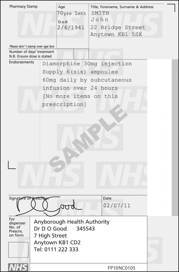
As the old saying goes, a picture is worth a thousand words, so here is an example of a Controlled Drug prescription on an FP10 prescription form.

Controlled drug prescription

Controlled drug prescription 1

* Sativex® (nabiximols) is an oromucosal spray extracted from Cannabis sativa, which is used in the treatment of moderate to severe multiple sclerosis spasticity.  Its main active components are tetrahydrocannabinol (THC) & cannabidiol (CBD).  Sativex® is a schedule 4, Part 1 drug; however, it is also listed as Class B.  This means that people can lawfully possess Sativex® if it was prescribed to them, but to redistribute to people without a prescription is a criminal act: distribution of a controlled substance.

References

Controlled drug prescription example – British National Formulary 

The post Controlled Drug Prescribing – OSCE guide appeared first on Geeky Medics.


Stoma examination – OSCE guide

$
0
0

Alongside a good abdominal examination it’s important to remember that many patients you encounter in hospital may already have abdominal pathology. Stomas are common things to come across and examination can be a skill frequently assessed in OSCEs. I know that my gut reaction (no pun intended) upon seeing stomas on examining a patients abdomen was usually to leave it well alone, but once you know what you’re doing this can be a simple (and actually very important) examination to have in your arsenal. If you aren’t really sure what a stoma is, a good place to start is with our summary of the different stoma types.

First of all, stomas are tougher than they look! A gentle examination of a patients stoma isn’t going to cause the bag to fall off or cause the patient any pain or discomfort; if these bags have to stay in place and not leak as the patient goes about their daily routine they are going to be able to withstand some gentle movement by medical students and in all honestly you won’t have to touch it that much anyway.

Introduction

Introduce yourself to the patient

Explain procedure and obtain consent

Wash hands

Expose patient adequately

Position patient supine at a 45 degree angle

Ask patient:
Do you have any pain in your tummy

Have you had any problems with your stoma

Inspection
Site

LIF: Colostomy

RIF: Ileostomy or urostomy


Number of lumens

1 and in RIF: End ileostomy or urostomy

1 and in LIF: End colostomy

2 joined and in RIF: Loop ileostomy

2 joined and in LIF: Loop colostomy


Spout

Spout present:

  • Ileostomy (contents toxic to skin)
  • Urostomy


No spout: Colostomy


Effluent (what’s coming out)

Hard stool – Colostomy

Soft stool – Ileostomy

Urine – Urostomy

Remember to feel the bag!


Surrounding skin quality

Any inflammation or excoriations?infection / poor stoma maintenance 


Any evidence of complications?

Haemorrhage – Peristomal skin inflammation

Parastomal hernia – Risk of bowel strangulation & necrosis

Prolapse – High output

Retraction – Obstruction

Auscultation

Listen below the umbilicus

Can bowel sounds be heard?

High pitched tinkling indicates obstruction.

To complete the examination…

Wash hands

Thank patient

Summarise findings

“Some Naughty Surgeons Never Stay in the Evenings and Like Porches”

  • Site
  • Number of lumens
  • Spout
  • Nature of effluent
  • State of surrounding skin
  • Evidence of complication
  • Likely type of stoma
  • Possible pathology/procedure


Example

This patient has a stoma in the LIF with one lumen and no spout. The effluent is solid faeces and the surrounding skin is intact with no evidence of inflammation. There is no evidence of complications. This is most likely an end colostomy. To complete my examination I would perform a full gastrointestinal exam.

References

1. Flesh and Bones of Surgery – page 58-59, 2007

2. 2nd edition Essential Examination –  page 104-106, 2010

The post Stoma examination – OSCE guide appeared first on Geeky Medics.

Chest X-ray (CXR) interpretation – a structured approach

$
0
0

Chest x-rays (CXR) are a frequently performed radiological investigation that you’ll be expected to be capable of interpreting (as due to the sheer volume of chest x-rays requested they are often not reported immediately). Therefore before hitting the wards as foundation doctors it is essential that you develop the ability to interpret chest x-rays, of particular importance is the ability to recognise findings that require immediate medical attention.

Confirm details

Always begin by checking the following:

  • Patient details (name / DOB)
  • Date & time the film was taken
  • Any previous imaging (useful for comparison)
Assess image quality

Then briefly assess the quality of the image: A mnemonic you may find useful is ‘RIPE’:

  • Rotation – the medial aspect of each clavicle should be equidistant from the spinous processes.
  • Inspiration – 5-6 anterior ribs, the lung apices, both costophrenic angles and lateral rib edges should be visible.
  • Projection – AP vs PA film.
  • Exposure – left hemidiaphragm visible to the spine and vertebrae visible behind heart.
CXR interpretation (ABCDE approach)
Airway

Trachea

Is the trachea significantly deviated?

  • The trachea is normally located centrally or just slightly off to the right
  • If the trachea is deviated, look for anything that could be pushing or pulling at the trachea.
  • Also inspect for any paratracheal masses / lymphadenopathy

Pushing of trachea – e.g. large pleural effusion / tension pneumothorax

Pulling of trachea – e.g. consolidation with lobar collapse

Rotation of the patient can give the appearance of a deviated trachea, so as mentioned above, check the clavicles to rule out rotation as the cause.

Pleural effusion with tracheal deviation 2

Pleural effusion with tracheal deviation 2


Carina and Bronchi

The carina is located at the point at which the trachea divides into the left and right main bronchus.

On a good quality CXR this division should be visible and is an important landmark when assessing nasogastric tube placement, as the NG tube should dissect the carina if it is correctly placed (i.e. not in the airway).

The right main bronchus is generally wider, shorter and more vertical than the left main bronchus. As a result it is more common for inhaled foreign objects to become lodged here (as the route is more direct). 

Depending on the quality of the CXR you may be able to see the main bronchi branching into further subdivisions of bronchi which supply each of the lungs lobes.

Carina & Bronchi (Normal CXR)

Carina & Bronchi (Normal CXR)


Hilar structures

  • The hilar consist of the main pulmonary vasculature and the major bronchi.
  • Each hilar also has a collection of lymph nodes which aren’t usually visible in healthy individuals.
  • The left hilum is often positioned slightly higher than the right, but there is a wide degree of variability between individuals.
  • The hilar are usually the same size, so asymmetry should raise suspicion of pathology.


Hilar enlargement can be caused by a number of different pathologies:

  • Bilateral symmetrical enlargement is typically associated with sarcoidosis.
  • Unilateral / asymmetrical enlargement may be due to underlying malignancy.

Abnormal hilar position can also be due to a range of different pathologies. You should (as with the trachea) look for any evidence of the hilar being pushed (e.g. by an enlarging soft tissue mass) or pulled (e.g. lobar collapse).


Breathing

Lung fields

Inspect the lung fields:

  • When looking at a CXR we divide each of the lungs into 3 zones, each occupying 1/3 of the height of the lung.
  • These zones do not equate to lung lobes (e.g. the left lung has 3 zones but only 2 lobes).
  • Inspect each of the zones of the lung first ensuring that lung markings occupy the entire zone.
  • Compare each zone between lungs, paying close attention for any asymmetry (some asymmetry is normal and caused by the presence of various anatomical structures e.g. the heart).
  • Some lung pathology causes symmetrical changes in the lung fields, which can make it more difficult to recognise, so it’s important to keep this in mind (e.g. pulmonary oedema).

Increased density in a given area of the lung field may suggest pathology (e.g. consolidation / malignant lesion).

The complete absence of lung markings within a segment of the lung field should raise suspicion of pneumothorax.

Lung tumour Right sided pneumonia1


Pleura

Inspect the pleura:

  • The pleura are not normally visible in healthy individuals.
  • Inspect the borders of each of the lungs to ensure lung markings extend all the way to the edges of the lung fields (if there appears to be an area lacking lung markings with decreased density this may suggest the presence of a pneumothorax).
  • Fluid (hydrothorax) or blood (haemothorax) can also accumulate in the pleural space, causing an area of increased opacity.

If a pneumothorax is suspected, you should reassess the trachea for evidence of deviation away from the pneumothorax which is in keeping with a tension pneumothorax. This is a medical emergency requiring immediate intervention. If  a tension pneumothorax is suspected clinically (shortness of breath and tracheal deviation) then immediate intervention should be performed without waiting for imaging as this condition will result in death if left untreated.  

Pleural thickening can be caused by mesothelioma.

Right sided pneumothorax Pleural thickening in the context of mesothelioma2
Cardiac

Assess heart size

In a healthy individual the heart should occupy no more than 50% of the thoracic width (e.g. a cardiothoracic ratio of <0.5).

This rule only applies to PA chest x-rays (as AP films exaggerate heart size), so you should not draw any conclusions about heart size from an AP film.

If the heart occupies more than 50% of the thoracic width (on a PA CXR) then this suggests abnormal enlargement (cardiomegaly). Cardiomegaly can occur for a wide variety of reasons including valvular disease, cardiomyopathy, pulmonary hypertension and pericardial effusion.


Assess heart borders

Inspect the borders of the heart which should be well defined in healthy individuals:

  • The right atrium makes up most of the right heart border.
  • The left ventricle makes up most of the left heart border.

The heart borders may become difficult to distinguish from the lung fields as a result of various pathological processes (e.g. consolidation) which cause increased opacity of the lung tissue.

  • Loss of definition of the right heart border is associated with right middle lobe consolidation
  • Loss of  definition of the left heart border is associated with lingular consolidation
Cardiomegaly

Cardiomegaly2

Diaphragm

The right hemi-diaphragm is in most cases higher than the left in healthy individuals (as a result of the underlying liver).  The stomach underlies the left hemi-diaphragm and is best identified by the gastric bubble located within it.

The diaphragm should be indistinguishable from the underlying liver in healthy individuals on an erect CXR, however if free gas is present as a result of bowel perforation air accumulates under the diaphragm causing it to lift and become visibly separate from the liver. If you see free gas under the diaphragm it indicates the need for urgent senior review as further imaging such as CT will likely be required to identify the source of free gas.

Pneumoperitoneum3

Pneumoperitoneum3


Costophrenic angles

The costophrenic angles are formed from the dome of each hemi-diaphragm and the lateral chest wall.

In a healthy individual the costo-phrenic angles should be clearly visible on a normal CXR as a well defined acute angle.

Loss of this acute angle (sometimes referred to as costophrenic blunting) can suggest the presence of fluid or consolidation in the area. Costophrenic blunting can also occur secondary to lung hyperinflation (seen in diseases such as COPD) as a result of diaphragmatic flattening and subsequent loss of the acute angle.

Costophrenic blunting secondary to pneumonia2

Costophrenic blunting secondary to pneumonia2

Everything else

Mediastinal contours

The mediastinum contains the heart, great vessels, lymphoid tissue and  a number of potential spaces where pathology can occur.  The exact boundaries of the mediastinum aren’t particularly visible on a CXR, however there are some important structures that you should assess.


Aortic knuckle:

  • Left lateral edge of the aorta as it arches back over the left main bronchus.
  • Loss of definition of the aortic knuckles contours can be caused by an aneurysm.


Aorto-pulmonary window:

  • The aorto-pulmonary window is a space located between the arch of the aorta and the pulmonary arteries.
  • This space can be lost as a result of mediastinal lymphadenopathy (e.g. malignancy).
Aortic knuckle & Aortopulmonary window

Aortic knuckle & Aortopulmonary window


Bones

Inspect the visible skeletal structures looking for any abnormalities (e.g. fractures / lytic lesions).


Soft tissues

Inspect the soft tissues for any obvious abnormalities (e.g. large haematoma).


Tubes / Valves / Pacemakers

Lines (e.g. central line / ECG cables).

Artificial valves (e.g. aortic valve replacement).

Pacemaker (often located below the left clavicle).

References
  1. By James Heilman, MD (Own work) [CC BY-SA 3.0 (http://creativecommons.org/licenses/by-sa/3.0) or GFDL (http://www.gnu.org/copyleft/fdl.html)], via Wikimedia Commons
  2. By James Heilman, MD – Own work, CC BY-SA 3.0, https://commons.wikimedia.org/w/index.php?curid=14634441
  3. By Clinical_Cases: I made the photo myself, licensed under Creative Commons license. – Modification of http://en.wikipedia.org/wiki/Image:Pneumoperitoneum.jpg Image source: http://clinicalcases.blogspot.com/2004/03/bloody-ascites-and-gas-under-diaphragm.html, CC BY-SA 2.5, https://commons.wikimedia.org/w/index.php?curid=2294165
  4. By Mikael Häggström, CC0, https://commons.wikimedia.org/w/index.php?curid=15228530

The post Chest X-ray (CXR) interpretation – a structured approach appeared first on Geeky Medics.

Nasal examination – OSCE guide

$
0
0

Nasal examination can sometimes appear in OSCEs. You’ll be expected to pick up the relevant clinical signs using your examination skills. Technique is very important in this station. This nasal examination OSCE guide provides a step by step approach to the station.

Introduction

Wash hands

Introduce yourself

Confirm patient details – name / DOB

Explain examination

Today I’d like to examine your nose, this will involve me having a feel of the outside of the nose and also gently looking inside the nose”

Gain consent Does everything I’ve said make sense?  Are you happy for me to go ahead?

Inspection

External

Look at the external surface of the nose noting:

  • Skin changes – e.g. skin lesions / erythema 
  • Deformity – inspect the nose from the front, side and standing behind the patient
    • Note any deviation in the nasal bones or cartilage

Internal

1. Ask the patient to look forwards, keeping their head in the neutral position.

2. Carefully elevate the tip of the nose with your thumb, so that the nasal cavity becomes visible. Use a pen torch or otoscope as a light source to externally illuminate the cavity.

3. Inspect the nasal mucosa for any abnormalities (including the septum).

4. Inspect and compare the nasal cavities alignment (note any septal deviation).


Further assessment

Further inspection can be done using an otoscope with a large speculum attached (inserting only the very tip into the nose), or using Thudicum’s speculum which essentially just widens the nasal cavity to allow you to peer in using a light source

Whichever method you use, you should inspect the various elements visible:

  • Nasal vestibule – skin changes (e.g. ulceration) / swelling / asymmetry
  • Nasal septum –  polyps / deviation
  • Inferior turbinates – asymmetry / inflammation / polyps

The turbinates are projections of bone, covered in nasal mucosa, that control airflow through the nose, exposing it to a large surface area of mucosa which both warms and cleans the air prior to it arriving at the lungs.

Palpation

Nasal bones and cartilage

Palpate the nasal bones assessing:

  • Alignment
  • Tenderness or irregularity (if suspicious of fracture in trauma)


Palpate the nasal cartilage assessing:

  • Alignment
  • Tenderness

Palpate the infraorbital ridges and assess eye movement if there is a history of trauma to screen for an orbital blowout fracture. The classical signs are of infraorbital tenderness, epistaxis and restricted eye movement (usually on vertical gaze).

Nasal airflow

There are two common methods via which to formally assess nasal airflow shown below.

Method 1

1. Place your thumb over the nostril not being assessed to occlude air flow.

2. Ask the patient to breath in through their nose and note the degree of airflow.

3. Repeat assessment on the other nostril, noting any difference in apparent airflow.

Reduce airflow through a particular nostril may indicate the presence of something blocking that air passage, such as a polyp or a deviated nasal septum.


Method 2

1. Place a cold shiny surface, such as a metal tongue depressor under the nose.

2. Observe for misting of the metal surface as the patient breathes, compare the misting pattern of the two nostrils.

Absence of misting, or a disparity in the amount of misting between the nostrils may suggest unequal or absent airflow through a particular nostril.

To complete the examination

Thank patient

Wash hands

Summarise findings


Suggest further assessments and investigations

  • Assessment of sense of smell
  • Regional lymph node examination
  • Flexible nasoendoscopy

The post Nasal examination – OSCE guide appeared first on Geeky Medics.

Certification of Death (UK) – OSCE guide

$
0
0

Completing a death certificate is a station in many-a final year OSCE, and more importantly is something that will need to be done in day-to-day life as a junior doctor. Knowing what to write where will speed this process up – great for the family and for you!

This guide will go through:

  • What to do when a patient has died and you are asked to do the death certificate
  • Some helpful tips and pointers
  • A copy of a blank Medical Certificate of Cause of Death form for you to print off and practice with
  • A few worked examples 🙂

What to do when a patient has died

  • Assess patient and confirm death, clearly documenting in the notes (see our guide here)
  • Discuss the death with the consultant in charge of the patient’s care (or a senior registrar) – this will help to clarify the cause to be written on the certificate.
  • Liaise with bereavement office to organise an appropriate time to attend with the patient’s medical notes to complete the death certificate and any other paperwork.
  • Complete Medical Certificate of Cause of Death form.
  • Perform a full external examination of the patient’s body in the mortuary (ensure no implantable devices present, check for bruising/pressure sores/any suspicious findings).

The Death Certificate

Click here to download a PDF copy of the blank death certificate here for use in the cases at the end of the guide.

Completing the death certificate

Personal details of the deceased

  • Age – you should record the age of the deceased in completed years or, if under one year, in completed months
  • Place of death – you should record to the best of your knowledge the precise place of death (e.g. the name of the hospital, or a private address)

Circumstances of certification

  • Last seen alive by me – record the date you last saw the deceased alive
  • Information from post-mortem – indicate if the cause of death takes into account information gained from a post-mortem or not
  • Seen after death – indicate who saw the deceased after death

Part I – Cause of death

  • Consider the main causal sequence of conditions that lead to death.
  • The disease or condition that led directly to death should be documented on the 1a line.
  • You should then work your way back through the other diseases that led to the eventual cause of death until you reach the underlying cause of death which initiated this chain of events. The lowest completed line in part 1 should therefore contain the underlying cause of death.
  • Some deaths may have only one condition that lead directly to death, such as a sub-arachnoid haemorrhage. In these cases it’s acceptable to complete only line 1a.
  • When stating the cause of death, be as specific as you are able to given the information you have. An example might be stating “Adenocarcinoma of the right main bronchus” rather than “Lung cancer”.
  • In some circumstances there can be 2 separate conditions that led directly to death and in these cases you should enter them both on the same line and then in brackets state that these are joint causes of death.

A few things that you CAN NOT write as a 1a:

  • “Failures” as a sole 1a – heart / kidney / liver / respiratory (Congestive Cardiac Failure is ok)
  • Asphyxia
  • Asthenia
  • Cachexia
  • Cardiac arrest
  • Coma
  • Exhaustion
  • Old Age/frailty as a sole cause of death (and this can only be used in those over 75)

Part II – Conditions that may have contributed to the death 

  • Part II allows you to document other conditions that were not part of the main causal sequence of death, but likely played a role in hastening the death.
  • An example might be ischaemic heart disease in a patient who died of pneumonia.
  • It is NOT somewhere where you should list the patient’s entire past medical history.

Other details at the bottom of the certificate

  • You should sign the death certificate and print your name in block capital letters beside it.
  • You should document your GMC number beside or under your name too
  • You should write down your GMC approved qualifications (often something like MBBS Medicine & Surgery)
  • Most commonly the residence is filled in as the hospital’s address (not your own personal address).
  • If the patient died in hospital, document the consultant’s name on the little line at the bottom of the certificate (easily missed).

Counterfoil

  • There is a counterfoil on the left hand side which gets left in the death certificate book
  • You need to document the patient’s name, the cause of death (Part 1) and conditions potentially contributing to death (Part 2).
  • You also need to document your personal details again.

Other Hints/Tips

  • _____ day of ______ refers to the “Fifth” day of “April 2015” (sounds simple, but I know someone who wrote “Tuesday” in an OSCE…).
  • Numbers should be written out in words
  • 1a should be the disease, illness or complication which led to death and not a mode of dying.
  • Circle the 1/2/3 & a/b/c with regards to post-mortem.

The back of the death certificate

There’s two boxes on the back of the death certificate that you may need to complete in certain circumstances:

  • Box A – If you have referred a death to the coroner put your initial here. It should be noted however that if you have simply discussed the case with the coroner and they have said no referral is required then you DO NOT need to complete this box.
  • Box B – If you may be in a position to provide more information about the cause of death in the future then you need to initial this box. This is the case when there are pending investigations not yet back or performed (e.g. histology, microbiology culture results, genetic analysis, post-mortem information). A request for this information will be sent to the consultant responsible for the patient

Reasons to refer to the Coroner…

  • Unknown cause of death
  • Sudden or unexpected death (inclusive of all deaths <24hours after admission to hospital)
  • Deceased person not seen by a doctor within 14 days before death
  • The death is considered suspicious/unnatural/violent
  • The death may be due to an accident, self-neglect or neglect on the behalf of others
  • The death is/could be due to the deceased’s prior employment (including industrial disease)
  • The death may be due to an abortion
  • The death occurred during an operation or before recovery from anaesthetic
  • The death occurred during or shortly after a period of police custody
  • The death may be suicide

These are (helpfully!) written on the back of the MCCD form.

How to refer to the Coroner

Coroners are often lawyers (and occasionally doctors, or dual-qualified). Talk to the Bereavement Team within the hospital with regards to getting in touch with the Coroner’s Office local to you.

Cases

Following on from this are some cases to work through with questions. You may want to print out a copy of the blank death certificate to fill out and treat it as a mock OSCE. Hidden in the tabs are the suggested answers.

Case 1

Mrs June Morbid was an 87 year old lady, whom you last attended to yesterday, on the ward round with the consultant (Dr Spot). She had suffered with advanced Parkinson’s disease and had been admitted 4 days previously with an aspiration pneumonia. This did not respond to antibiotic treatment, and the decision to palliate was made by the consultant after discussion with the family. Mrs Morbid peacefully passed away last night with her family around her, and her death was verified by your colleague on the night shift. You have been asked to fill in the death certificate after your ward round.

Completed death certificate

Completed death certificate

Case 2

Mr Clive Matchstick (86 years old) was admitted to the ward a week ago from a local nursing home having vomited. Whilst in hospital he developed urinary incontinence and sepsis. He was treated for urosepsis, but unfortunately passed away. You confirmed his death this morning and had reviewed him last night before going home with his consultant Dr Johnson . He has a past history of: ischaemic heart disease,  type 2 diabetes mellitus, Charcot’s deformity on the left foot and an amputation of the big toe on the right.

Completed death certificate

Completed death certificate

Case 3

Mr Samuel Clock (75 years old) had been an in-patient on the ward you are working on for 2 weeks. He was being treated for a community-acquired pneumonia which was a CURB-65 score of 4. His condition had progressively worsened when you reviewed him with his consultant Dr Tyvand last night and the decision was made to switch to a palliative approach of management. He passed away this morning, with his wife by his side. You confirmed his death on the ward. He has a past medical history of ischemic heart disease, hypertension, mesothelioma, type 2 diabetes and benign prostate hypertrophy.

Completed death certificate

This is a trick question (cheeky, I know)…

This is a situation where the medical practitioner should have a conversation with the Coroner prior to issuing any certificate of cause of death as a post-mortem will most likely be required. Mesothelioma is a is almost always attributed to asbestos exposure and therefore falls into category of disease related to occupation which may have contributed to the death. The damage most often occurs 20 – 60 years after asbestos exposure. It would be very unlikely that you would issue a certificate, but if the Coroner instructed that you could do so, it might look a bit like this.

Completed death certificate

References

  1.  Southampton University Hospitals NHS Trust – Medical Certificate of Cause of Death – Notes for doctors – LINK

The post Certification of Death (UK) – OSCE guide appeared first on Geeky Medics.

Intramuscular injection (IM) – OSCE guide

$
0
0

Intramuscular (IM) injections administer medication deep into the muscle tissue, enabling faster absorption and larger systemic doses than subcutaneous injections. This guide discusses how to perform an IM injection in an artificial OSCE setting and should not be used as a guide to administering injections to actual patients without first consulting your local medical school or hospital guidelines. Check out the Intramuscular (IM) injection OSCE mark scheme here.

Introduction

Introduce yourself

Wash hands

Confirm the patient’s details

Explain the procedure

Check the patient’s understanding of the medication being given, and explain the indication for the medication.

Check for allergies

Check if the patient has a bleeding disorder or takes anticoagulant medication (possible contraindications).

Gain consent to proceed

Gather equipment

  • Equipment tray
  • The medication to be administered
  • Patient’s prescription
  • Syringe – the smallest syringe that will accommodate the medication volume
  • Injecting needle – (21–23 gauge) – 25mm in length is standard 
  • Drawing up needle 
  • Antiseptic swab –  70% isopropyl alcohol wipe
  • Gauze or cotton swab
  • Sharps container (for disposal of the needle)
  • Apron
  • Non-sterile gloves

7 Rights

Return to the patient. Before proceeding, check the 7 rights of medication administration

1. Right person – check the patient’s arm band against the name on the prescription. Where possible aim to use 2 identifiers (e.g. from the patient and the arm band)

2. Right drug – check the labelled drug against the prescription – ensure expiry date is appropriate

3. Right dose – check the dose against the prescription

4. Right time – confirm when the last dose was given

5. Right route

6. Right to refuse – has the patient consented?

7. Right documentation of prescription and allergies – does the patient have any allergies?

Once these have been confirmed prepare the medication. Always use a separate drawing up needle and injection needle.

Injection steps

1. Wash hands.

2. Don gloves and apron.

3. Draw up the appropriate medication into the syringe using a drawing needle.

4. Remove the drawing needle and attach the needle to be used for injection.

5. Choose an appropriate site. Common sites include:

  • Deltoid (upper arm)
  • Ventrogluteal (upper outer buttock) – ideal for larger volumes
  • Vastus lateralis (anterior lateral thigh)
  • Do NOT use a site that is inflamed, irritated, bruised or contains scar tissue.

See the end of the guide for further information regarding the most commonly used sites.

If multiple injections are given, use different sites for each subsequent injection. If frequent injections are given, rotate sites.

6. Position the patient to provide optimal access to your chosen site.

7. Swab the site with an antiseptic swab and wait until it is dry.

8. Gently place traction on the skin with your non-dominant hand away from the injection site, continuing the traction until the needle has been removed from the skin. This is known as the Z track technique (see below).

9. Warn the patient of a sharp scratch.

10. Holding the syringe like a dart in your dominant hand, pierce the skin at a 75 – 90 degree angle. Insert the needle quickly and firmly, with the bevel facing upwards, leaving approximately 1/3 of the shaft exposed (however this varies between sites and patients).

11. Aspirate to check the location of the needle. If blood appears, remove the syringe and prepare a new injection (explaining what occurred to the patient).

12. If no blood appears on aspiration, inject the contents of the syringe while holding the barrel firmly. Inject the medication slowly at a rate of approximately 1ml every 10 seconds.

13. Remove the needle and immediately dispose of it appropriately (into a sharps container).

14. Release the traction on the skin.

15. Apply gentle pressure over the injection site with a cotton swab or gauze.  Do NOT rub the site.

Z – track technique – releasing the traction on the skin changes the alignment of the subcutaneous and muscle tissue layers, ‘locking’ the medication into the muscle layer.

To complete the procedure

Replace cotton swab with a plaster.

Thank the patient

Discuss post injection care:

  • Warn them that the injection site may be sore for one or two days, but this is normal.
  • Other potential complications include: haematoma, persistent nodules, local irritation (and rarely anaphylaxis).
  • Advise the patient to watch for a developing rash, breathing difficulty or other relevant concerning symptoms. They should discuss this with a doctor if concerned.

Wash hands

Document that the medication has been given on the medication chart and in the patient’s notes.

Have the patient observed for 15 – 30 minutes for adverse effects.

Injection sites

Below are some more details surrounding the common sites used for IM injections, however these are brief notes and therefore should NOT be relied upon in isolation for carrying out IM injections, instead you should consult your local medical school or hospital guidelines.

Deltoid site

Position of patient

  • Have the patient sitting down, with their entire shoulder and arm exposed.
  • Position their elbow flexed and ask them to relax.

Site of injection

  • Palpate the lower edge of the acromial process and inject approximately 2.5cm below this.

Ventrogluteal site

In the past the dorsogluteal site was very popular, however due to potential complications such as sciatic nerve injury or superior gluteal artery injury it is now not recommended. Instead the ventrogluteal site is used as a safer alternative as it avoids all major nerves and blood vessels.

Position of patient

  • The patient can be prone, semi-prone or supine for this procedure, so choose whichever is most comfortable for the patient.

Site of injection

  • Place palm of your right hand over the greater trochanter of the patients left hip (or vice versa).
  • Extend your index finger to touch anterior superior iliac crest.
  • Then stretch your middle finger to form a V (thumb pointing towards the front of leg)
  • Insert the needle into the V at 90 ̊

References

Click to show
  • Department of Health. (2014). Intramuscular Injections. Government of Western Australia. Retrieved from: http://www.kemh.health.wa.gov.au/development/manuals/O&G_guidelines/sectiona/1/a1.10.pdf
  • Department of Health. (2016) The Australian Immunisation Handbook – Administration of vaccines. Ed. 10 The Australian Government. Retrieved from: http://www.health.gov.au/internet/immunise/publishing.nsf/content/Handbook10-home~handbook10part2~handbook10-2-2#2-2-5
  • World Health Organisation – WHO Best Practices for Injections and Related Procedures Toolkit. Retrieved from: http://apps.who.int/iris/bitstream/10665/44298/1/9789241599252_eng.pdf

The post Intramuscular injection (IM) – OSCE guide appeared first on Geeky Medics.

Ankle-brachial pressure index (ABPI) measurement – OSCE guide

$
0
0

The ankle-brachial pressure index (ABPI) is a non-invasive method of assessing the extent of chronic peripheral arterial disease in the lower limbs.  It is a ratio composed of the blood pressure in the brachial artery and the pressures in the foot arteries (dorsalis pedis and the posterior tibial artery). This guide provides a step by step approach to performing ABPI measurement in an OSCE setting.

Introduction

Wash hands

Introduce yourself – state your name and role

Confirm patient details – name / DOB

Explain the procedure:

  • “I would like to measure the pressures in your arm and ankle”
  • “This will involve inflating a cuff around your arm and ankle briefly and listening to your pulse with this probe”
  • “It shouldn’t be painful, but it may feel a little tight temporarily, please let me know if you want to stop at any point”

Check understanding: “Do you understand everything I’ve mentioned? Do you have any questions?”

Check if the patient currently has any pain: “Do you have any pain anywhere?”

Rule out diabetes: “Do you suffer from diabetes?” (see reasoning below)

Gain consent: “Are you happy for me to continue with the assessment?”

Calcified vessels in diabetes sufferers will lead to artificially high ABPI values due to vessels which are not compressible. In such cases measure the Toe pressure (with a special toe cuff) rather than the ankle pressure. This is called the Toe Brachial Pressure Index (TBPI).

If the patient has leg ulcers these can be covered in sterile cling-film and the cuff applied as above, provided the patient doesn’t feel discomfort

Gather equipment

  • Sphygmomanometer
  • Doppler probe
  • Ultrasound gel

Check probe is functioning correctly (battery / sound volume)

Measure ABPI

Measuring the brachial pressure

1. The patient must be lying supine for the measurements on an examination couch.

2. Place the sphygmomanometer cuff over the left arm proximal to the brachial artery (avoiding any cannulas) and position the doppler probe on the brachial artery at a 45° angle (medial to the biceps tendon at the antecubital fossa).

3. Inflate the cuff 20-30 mmHg above the pressure at which the doppler pulse is no longer heard. Deflate the cuff slowly and note the pressure at which you first detect a pulse from the doppler. This is the systolic pressure in that vessel.

4. Now repeat this process on the right brachial artery.

5. The higher of the two pulses will be used as part of the ratio.

Measuring the ankle pressure

1. Place the sphygmomanometer on the left ankle and position the doppler probe over the posterior tibial artery (behind the medial malleolus of the foot). Measure the pressure in the same way as for the brachial artery: inflate and deflate the cuff slowly to detect the pressure at which blood returns to the posterior tibial artery.

2. Repeat this process (keep the sphygmomanometer in the same place) at the dorsalis paedis artery of the left foot (lateral to the extensor hallucis longus tendon).

3. Use the highest of the two pressures obtained from the posterior tibial artery (PTA) and dorsalis pedis (DP) for the ratio of the left ABPI.

4. Repeat the same process on the right ankle to work out the ratio for the right ABPI.

To complete the examination…

Thank the patient

Wash hands

Record left and right ABPI in the patient’s notes


Suggest further assessments and investigations


Examiner question: “If you find an abnormal ABPI result, what would be the next steps in investigation and management?”

Answer: If mild disease is present, this may be managed conservatively. If further imaging is needed, then a duplex ultrasound of the lower limb arteries can be undertaken. Prior to surgical or endovascular interventions a CT or MR angiogram may be needed.“

Summary

Left ABPI(Highest pressure of either left PTA or DP) ÷ (Highest brachial pressure)

Right ABPI = (Highest pressure of either right PTA or DP) ÷ (Highest brachial pressure)

Example

  • Right brachial artery: 120 mmHg
  • Left brachial artery: 125 mmHg
  • Right DP: 80 mmHg
  • Right TP: 75 mmHg


Right ABPI = 75/125 = 0.6


Interpret results

ABPI Meaning
>1.2 Abnormally hard vessel (e.g. calcified) – this can often be a false negative as there is likely significant peripheral vascular disease but the hardened vessels give a higher ABPI reading, so correlation with clinical findings is advised.
1.0-1.2 Normal
0.8-0.9 Mild arterial disease: mild claudication
0.5-0.79 Moderate arterial disease: severe claudication
<0.5 Severe arterial disease: rest pain, ulceration and gangrene (critical ischaemia)


Errors can occur due to:

  • Irregular pulse (AF)
  • Cuff positioned incorrectly
  • Calcified vessels (diabetes)

References

Click to show

[1]. Mo Al-Qaisi, David M Nott, David H King, and Sam Kaddoura. (2009). Ankle Brachial Pressure Index (ABPI): An update for practitioners. Vasc Health Risk Manag. 5, 833–841.


[2]. McDermott MM, Criqui MH, Liu K, Guralnik JM, Greenland P, Martin GJ, Pearce W (2000). “Lower ankle/brachial index, as calculated by averaging the dorsalis pedis and posterior tibial arterial pressures, and association with leg functioning in peripheral arterial disease”. J Vasc Surg. 32 (6): 1164–71


[3]. Gogalniceanu P, Pegrum J, Lynn W. (2015) Physical Examination for Surgeons – a guide to the MRCS OSCE. Cambridge University Press, UK.

CONTENT REVIEWED BY

Dr Peter Gogalniceanu – Vascular surgeon 

The post Ankle-brachial pressure index (ABPI) measurement – OSCE guide appeared first on Geeky Medics.

Ankle and Foot examination

$
0
0

Ankle and foot examination can occasionally appear in OSCEs, so it’s important you’re familiar with it. Check out the ankle and foot examination mark scheme here.

Introduction

Wash hands

Introduce yourself – state your name and role

Confirm patient details – name and DOB


Explain examination:

“I’d like to examine your ankles and feet. This will involve having a look and feel of these joints, in addition to assessing the joints’ movement”


Gain consent – “Do you understand everything I’ve said?” “Are you happy to go ahead with the exam?”

Gain adequate exposure – both legs should be exposed from the knee down

Position patient – ask patient to stand up straight with their feet aligned facing forwards

Look

Gait

Is the patient demonstrating a normal heel strike / toe off gait?

Is each step of normal height?increased stepping height is noted in foot drop

Is the gait smooth and symmetrical?

Examine the patient’s shoes – evidence of asymmetrical wearing may indicate abnormal gait 

Inspect from the front

Symmetry of feet and ankles

Toe alignment – hallux valgus of the big toe may be noted

Bunions – located at the 1st metatarsal phalangeal joint (MTP) joint

Toe clawing?

Scarssuggestive of previous injury / surgery

Calluses – may indicate foot / gait deformity or poorly fitting footwear

Swelling or erythema of the foot or ankle – may suggest injury / inflammatory arthritis / infection


Inspect from the side

Foot arches – observe for evidence of flat feet (pes planus) or high arched feet (pes cavus)

If patient has flat feet ask to stand on tiptoes – supple flat feet will correct / rigid flat feet will not


Inspect from the back

Foot / ankle symmetry – heel alignment – valgus or varus deformity?

Achilles tendon – any obvious deformity / discontinuity / erythema?

Feel

Ask the patient to lay on a bed

Assess temperature and compare between legs – ↑ temperature may indicate inflammatory pathology

Assess pulses in both feet – posterior tibial and dorsalis pedis 


Palpate the achilles tendon:

  • Position the patient kneeling on a chair
  • Palpate the gastrocnemius muscle and achilles tendon
  • Note any focal tenderness, swelling or gaps in the tendon (suggestive of rupture)


Palpate the joints and bones

Work distal to proximal – assess for tenderness / swelling / irregularity  

  • Squeeze MTP joints – observe patient’s face for discomfort
  • Tarsal joint
  • Ankle joint 
  • Subtalar joint
  • Medial / lateral malleoli
  • Proximal fibula

Move

Active and passive assessment

Assess each of the following movements actively and passively (feeling for crepitus)

Foot plantarflexion – push your feet downwards, like pushing a car pedal” – 30-40 º

Foot dorsiflexion – “point your feet towards your head” – 12-18 º

Toe flexion – “curl up your toes”

Toe extension – “point your toes towards your head”

Foot inversion

Foot eversion

Passive assessment only

Assess each of the following movements passively

Midtarsal joints – hold ankle with one hand whilst moving the forefoot through dorsiflexion, plantarflexion, adduction, abduction, supination and pronation. 


Special tests

Simmonds’ test

Simmonds’ test is used to assess for rupture of the achilles tendon

1. Ask patient to kneel on a chair with their feet hanging off the edge

2. Squeeze each calve in turn

3. Normally the foot should plantarflex

4. If the achilles tendon is ruptured there will be no movement of the foot

To complete the examination

Thank patient

Wash hands

Suggest further assessments and investigations

  • Examine the knee and hip joint 
  • Full neurovascular examination of the lower limbs
  • Further imaging of the relevant joints if indicated (Xray / CT / MRI)

The post Ankle and Foot examination appeared first on Geeky Medics.


Testicular examination – OSCE guide

$
0
0

Testicular examination can occasionally appear in OSCEs and if it does you need to have practiced the routine to look slick and professional. This testicular examination OSCE guide provides a structured approach to examining the testicles. This is an intimate examination and therefore extra attention should be paid to the communication aspect to ensure the patient feels as comfortable as possible. Check out the testicular examination OSCE mark scheme here.

Introduction

Wash hands

Introduce yourself – name / role

Confirm patient details – name / DOB


Explain examination:

I need to carry out an examination of your testicles, this will involve firstly inspecting the testicles and surrounding area, then I will examine the testicles

I’m required to have a chaperone present, this will most likely be a nurse from the ward, are you ok with that?


Gain consent :

Do you understand everything I’ve said?  

Are you happy for me to examine you?


Get a chaperone – this is absolutely essential and often carries significant marks in the OSCE

Position patient – examination of the testicles is best performed with the patient standing

Expose patient – exposure should be from the waist down

Don gloves 

Inspection

General inspection

Inspect the patient’s genital region and the surrounding areas (i.e. penis / groin / lower abdomen):

  • Skin changes – rash / bruising / swelling
  • Scars
  • Obvious masses


Inspection of the scrotum

Ask the patient to hold their penis out of the way to allow easier inspection of the scrotum.

Inspect the scrotum from the front, sides and the posterior aspect by lifting the scrotum.


Inspect the scrotum for the following…

  • Skin changes – rash / ulcers / erythema (e.g. cellulitis / fungal infection)
  • Scars – may provide clues as to previous operations (e.g. vasectomy)
  • Masses – note any obvious lumps, these will require examination later
  • Swelling – unilateral or bilateral? / associated with erythema? 
  • Bruising
  • Necrotic looking tissue – Fournier’s gangrene is a diagnosis not to be missed!

Palpation

Examine each testicle individually.

If abnormalities have been identified on inspection or the patient is concerned about a particular testicle, start examination on the other testicle.

Ask the patient to report any pain or discomfort they experience during the examination.

Testes

Use your thumb and index finger to gently palpate the whole testicle.

If you are unable to locate a testicle, palpate along the path of the inguinal ligament for an undescended testicle (if the patient also has a scar this would suggest a previous orchidectomy)

If a mass is found assess the following…

  • Size / shape
  • Regularity – regular vs irregular
  • Consistency – hard (solid) / soft (cystic) / “Bag of worms” (varicocele) 
  • Discomfort – try to identify the specific area causing pain
  • Are you able to get above the mass?- No = inguinal scrotal hernia


Is the mass fixed to the testicle or separate?

  • Separate + hard (solid) = epididymitis / orchitis
  • Separate + cystic (soft / fluctuant) = epidermal cyst / spermatocele


Is there a cough impulse? – presence of a cough impulse suggests hernia / varicocele

Does the mass transilluminate? – transillumination suggests the mass is fluid filled – e.g. hydrocele

Epididymis

Palpate the epididymis (located at the posterior aspect of the testicle).

Pain in the epididymis may suggest epididymitis.


Phren’s test

If testicular pain is relieved by elevating the testes this is strongly suggestive of epididymitis.


Spermatic cord

Start palpation at the superior aspect of the testicle using your thumb and index finger.

The spermatic cord should be palpable connecting to the testicle at this region.

Palpate along the cord assessing for masses and tenderness.


Lymph nodes

Assess the local lymph nodes in the inguinal region for evidence of lymphadenopathy (scrotal infection).

To complete the examination

Thank patient

Allow patient time to get dressed

Dispose of gloves

Wash hands


Summarise findings 

Today I performed a testicular examination on John Doe, a 42 year old gentleman. On inspection there were no abnormalities identified, however on palpation there was a 1cm smooth solid mass noted on the left testicle. The mass was non tender and fixed to the underlying testicle. I was able to get above the mass and there was no cough impulse or transillumination.  Some lymphadenopathy was noted in the left inguinal region


Suggest further assessments and investigations

The post Testicular examination – OSCE guide appeared first on Geeky Medics.

Hearing assessment and otoscopy – OSCE guide

$
0
0

Hearing assessment and otoscopy frequently appear in OSCEs. You’ll be expected to pick up the relevant clinical signs using your examination skills. Technique is very important in this station, so ensure you’ve practiced how to hold and use an otoscope before your exam. This guide provides a clear step by step approach to the station. Check out the Hearing assessment and Otoscopy OSCE mark scheme here.

Introduction

Wash hands

Introduce yourself

Confirm patient details – name / DOB

Explain examination

Today I’d like to examine your ears, this will involve me having a look inside your ears using a special piece of equipment known as an otoscope. In addition I’ll also be assessing your hearing using a number of different tests

Gain consent Does everything I’ve said make sense?  Are you happy for me to go ahead?

Gross hearing assessment

Ask the patient if they have noticed any change in their hearing recently.

Explain that you’re going to say a word or number and you’d like them to repeat it back to you.

1. With your mouth approximately 15cm from the ear, whisper a number or word.

2. Mask the ear not being tested by rubbing the tragus.

3. Ask the patient to repeat the number or word back to you.

4. If the patient repeats the correct word or number, repeat the test at an arm’s length from the ear (normal hearing allows whispers to be perceived at 60cm).

5. Assess the other ear in the same way.

Weber’s test

1. Tap a  512 Hz tuning fork and place in the midline of the forehead

2. Ask the patient Where do you hear the sound?

  • Normal – sound is heard equally in both ears
  • Neural deafness – sound is heard louder on the side of the intact ear
  • Conductive deafness – sound is heard louder on the side of the affected ear

Rinne’s test

1. Tap a 512 Hz tuning fork and place at the external auditory meatus (EAM). Ask the patient if they are able to hear it (air conduction).

2. Now move the tuning fork (whilst still vibrating), placing its base onto the mastoid process (bone conduction).

3. Ask the patient if the sound is louder in front of the ear (EAM) or behind it (mastoid process)

  • Normal – Air conduction > Bone conduction (Rinne’s positive)
  • Neural deafness  Air conduction > Bone conduction (both air and bone conduction ↓ equally)
  • Conductive deafness – Bone conduction > Air conduction (Rinne’s negative)

Otoscopy

Ask the patient if they have any ear discomfort (if so examine the non-painful side first).

Pinnae

Inspect the pinnae –  note any deformity / ear piercings

Inspect behind the pinnae – skin changes / erythema

Ear canal / tympanic membrane

Ensure the light is working on the otoscope and apply a sterile speculum (the largest that will comfortably fit in the external auditory meatus).

1. Pull the pinna upwards and backwards – to straighten the external auditory meatus

2. Position otoscope at the external auditory meatus:

  • Otoscope should be held in your right hand for the patient’s right ear and vice versa
  • Hold the otoscope like a pencil and rest your hand against the patient’s cheek for stability

3. Advance the otoscope under direct vision

4. Look for any wax, swelling, erythema, discharge or foreign bodies

5. Examine the tympanic membrane:

  • Colour pearly grey and translucent (normal) / erythematous (inflammation)
  • Erythema or bulging of the membrane? inspect for a fluid level e.g. otitis media
  • Perforation of the membrane? note the size of the perforation
  • Light reflex present? – absence / distortion may indicate ↑ inner ear pressure e.g. otitis media 
  • Scarring of the membrane? – tympanosclerosis – can result in significant hearing loss


6. Withdraw the otoscope carefully

7. Discard the otoscope speculum into a clinical waste bin

To complete the examination

Thank patient

Wash hands

Summarise findings


Suggest further assessments and investigations

The post Hearing assessment and otoscopy – OSCE guide appeared first on Geeky Medics.

Urinalysis – OSCE guide

$
0
0

Urinalysis (urine dipstick testing) frequently appears as an OSCE station, so you need to get familiar with the process to look smooth in the exam. It’s actually a very simple process, but people often fail to revise this station and lose marks unnecessarily. With a little practice, you can ensure you get full marks every time! Check out the urinalysis OSCE mark scheme here.

Gather equipment

  • Alcohol gel
  • Gloves
  • Apron
  • Urine dipsticks
  • Urine sample
  • Paper towels
  • Gather equipment

Inspect urine

1. Wash hands, don gloves and apron

2. Confirm patient details are correct on the sample bottle – name / DOB / hospital number

3. Inspect the colour of the urine:

  • Straw coloured – normal
  • Dark concentrated urine dehydration
  • Red macroscopic haematuria / rifampicin / porphyria / beetroot
  • Brown – bile pigments / myoglobin / antimalarials 

4. Inspect the clarity of the urine:

  • Clear normal
  • Cloudy / debris  urinary tract infection (UTI)
  • Frothy – nephrotic syndrome

5. Remove the sample bottles’ cap and assess urine odour:

  • Offensive urine UTI
  • Sweet – glycosuria
  • Wash hands

Perform dipstick testing

1. Check urine dipsticks’ expiry date

2. Remove a testing strip from the container (avoiding touching the testing zones)

3. Insert test strip into urine sample (ensuring all test zones are immersed)

4. Remove the strip, ensuring to tap off residual urine before removing from the sample bottle

5. Ensure test strip remains in a horizontal orientation (to avoid cross contamination of testing zones)

6. Use the dipstick analysis guide on the side of the testing strip container to interpret the findings

7. Different tests on the strip are required to be read at different times, so ensure you interpret the appropriate test at the correct time interval – e.g. 60 seconds for protein

8. Once you have interpreted all of the tests, discard the strip into the clinical waste bin along with your gloves and apron

9. Wash hands

  • Check urine dipstick expiry date

To complete the procedure

Summarise findings

Document urinalysis results

Indicate that depending on the results, further investigations may be required:

  • ↑ WCC / Leukocytes – ?UTI –  send urine for culture (MSU/CSU)
  • ↑ Glucose – ?Diabetes  capillary blood glucose

Urine dipstick tests explained

pH – indicates acidity of urine – e.g. ↓pH in systemic acidosis

Specific gravity – indicates amount of solute dissolved in urine – ↓ in diabetes insipidus

Blood  – indicates number of red blood cells in urine – ↑ in haematuria

Protein – indicates level of protein in the urine – ↑ nephrotic syndrome

Leukocyte esterase – enzyme produced by neutrophils (WCC in urine) – ↑ in UTI

Nitrites – breakdown products caused by Gram -ve organisms – Gram -ve UTI e.g. Ecoli

Ketones – breakdown product of fatty acid metabolism – ↑ starvation / ↑DKA

Glucose – ↑ hyperglycaemia e.g. poorly controlled diabetes

Bilirubin – Indicates ↑ conjugated bilirubin (water soluble) – ↑ biliary tract obstruction

Urobilinogen – if raised indicates ↑ bilirubin turnover – ↑malaria / ↑haemolytic anaemia 

The post Urinalysis – OSCE guide appeared first on Geeky Medics.

Inhaler technique – OSCE guide

$
0
0

Inhaler technique is a common topic in information giving OSCE scenarios. This guide demonstrates a structured approach to explaining inhaler technique in an OSCE setting. Check out the inhaler technique mark scheme here.

Introduction

Introduce yourself

Wash hands

Confirm patient details – name / DOB

Check patient’s understanding of their inhaler – allowing you to tailor your explanation to the patient’s level of knowledge

Explanation

Explain what the inhaler device is…

“You have been started on ….(name of inhaler)…for your asthma/COPDshow the patient the inhaler device

Explain when the inhaler device should be used…

Preventer (e.g. beclomethasone inhaler)

(Name of inhaler) is a preventer – it helps to reduce the swelling in the airways and stops them from being so sensitive. You use this to lower the risk of severe attacks. I would like you to inhale …(x puff(s))…(x time(s) a day)…everyday. It’s really important that you don’t miss doses, as regular use is key to keeping your asthma/COPD under control  Remind the patient to rinse mouth after use if the inhaler contains a steroid due to risk of oral candidiasis.


Reliever (e.g. salbutamol inhaler)

(Name of inhaler) is a reliever. This is useful to help relieve immediate wheezing/asthma attacks. It works by relaxing the airways so that you can breathe more easily. You shouldn’t need this more than 3 times a week if your asthma is well controlled. Ask your GP for a review if you are using this more frequently. I would like you to inhale (x puff(s)) when you feel short of breath.

If prescribing SMART (Symbicort Maintenance and Reliever Therapy) regime

Symbicort is used as both a preventer and a reliever. You need to use this regularly …(x puff(s))…twice a day to prevent symptoms and …(x puff(s))…each time you have an attack. Remind the patient to rinse mouth after use due to risk of oral candidiasis. 

Show patient the dose counter on the inhaler where applicable.  Ask the patient to summarise the key points back to you to demonstrate understanding.

Demonstration

Explain the steps below as you demonstrate

1. Prepare the inhaler (take off the lid / shake if MDI / insert capsule if handihaler)

2. Load the dose – (press button to puncture capsule if handihaler  press lever once if accuhaler/ twist bottom if turbohaler)

3. Breathe out gently as far as is comfortable

4. Tightly seal lips around the mouthpiece

5. Breathe in:

  • Dry powder inhalers (DPI) need to be breathed in quick and deep
  • Metered dose inhalers (MDI) need to be breathed in slow and deep
  • Soft mist inhalers (SMI) need to be breathed in slow and deep

6. Remove inhaler from mouth, hold breath for as long as is comfortable

7. Repeat procedure as directed

Assess inhaler technique

Ask the patient to carry out the procedure themselves whilst you observe.

Most patients’ techniques will require tweaking.

Point out the positives …You are doing X&Y very well… then introduce room for improvement …”but doing A&B may help your inhalers work more effectively for you

DEMONSTRATE > OBSERVE > FINE TUNE > REPEAT AS NECESSARY 

Spacer devices

Spacers are used to improve drug deposition to the lungs in patients who cannot master their aerosol inhaler technique. They are useful in reducing side effects of high dose inhaled corticosteroids by reducing the amount of drug deposited in the mouth. Commonly used spacers are Volumatic and AeroChamber.

1. Prepare inhaler (shake aerosol inhaler)

2. Attach inhaler mouthpiece to the spacer device

3. Breathe out gently as far as is comfortable

4. Seal lips around the spacer mouthpiece

5. Release 1 dose into the spacer device

6. Breathe in and out through the spacer mouthpiece several times

7. Administer second dose if needed and finish

The spacer device should be washed with detergent (washing up liquid is fine) once a month and left to air-dry. It should never be wiped dry as this can cause static within the device and drug particles will stick to sides of the spacer as a result. Spacers should be replaced at least once a year.

To close the consultation

Ask if the patient has any questions or concerns

Provide information leaflet if available

Advise the patient to get in touch should they have any more questions or concerns

Thank patient

Wash hands

Commonly used inhalers

TinyGrab Screenie at 2015-04-09 01-37-51

The post Inhaler technique – OSCE guide appeared first on Geeky Medics.

Controlled Drug Prescribing – OSCE guide

$
0
0

There are special prescription requirements for certain drugs, classed as Controlled Drugs.  These requirements are in part to monitor the distribution of drugs that are potentially harmful when misused, to prevent patients developing a drug dependency, and to prevent those who misuse drugs from obtaining a prescription by deception.

Controlled drug prescribing is a common station to find in final-year OSCEs and you’ll be writing a lot of these as a doctor, so it’s worth learning well.  Controlled drug (CD) prescriptions aren’t too different from normal prescriptions, so, as always, getting good at the basics is advisable!

Acts and regulations

The Misuse of Drugs Act 1971 splits controlled drugs into classes A, B and C depending on the harmfulness attributable to the drug when it is misused. This classification forms the basis for legal penalties for unlawful possession and distribution.

The Misuse of Drugs Regulations 2001 sets out regulations surrounding controlled drugs, including who can supply and possess controlled drugs in a professional capacity. These regulations split controlled drugs into schedules:

  • Schedule 1 includes drugs like cannabis*, which are never used for medical purposes.
  • Schedule 2 includes drugs such as diamorphine, morphine, pethidine and amphetamine.
  • Schedule 3 includes drugs such as barbiturates, buprenorphine, midazolam and temazepam.
  • Schedules 4 and 5 exist, but the drugs within are not subject to special prescription requirements.

All drugs in schedules 2 and 3 (other than temazepam) are subject to prescription requirements. You do not need to write a controlled drug prescription for temazepam.

Requirements for a controlled drug prescription

Controlled drug prescriptions must:

  • Be indelible
  • Be dated
  • Be signed by the prescriber
  • Include the prescriber’s address
  • Include the name and address of the patient
  • Include the date of birth of the patient (and age if <12 years)
  • Include the form of the preparation –  e.g. tablets, even if there is only one form of the drug
  • Include the strength of the preparation, if appropriate
  • Include the total quantity in both words and figures OR number in words and figures of doses to be supplied
  • Include the dose to be taken


Controlled drug prescriptions should:

  • Include the patient identifier (NHS number in England, Community Health Index number in Scotland, there is no guidance for Wales or Northern Ireland)
  • Include the prescriber’s GMC number
  • Have a diagonal line drawn underneath the prescription to indicate no more items, OR have “No more items” written under the last prescription

As the old saying goes, a picture is worth a thousand words, so here is an example of a controlled drug prescription on an FP10 prescription form.

Controlled drug prescription

Controlled drug prescription 1

* Sativex® (nabiximols) is an oromucosal spray extracted from Cannabis sativa, which is used in the treatment of moderate to severe multiple sclerosis spasticity.  Its main active components are tetrahydrocannabinol (THC) & cannabidiol (CBD).  Sativex® is a schedule 4, Part 1 drug; however, it is also listed as Class B.  This means that people can lawfully possess Sativex® if it was prescribed to them, but to redistribute to people without a prescription is a criminal act: distribution of a controlled substance.

References

Controlled drug prescription example – British National Formulary 

The post Controlled Drug Prescribing – OSCE guide appeared first on Geeky Medics.

Viewing all 223 articles
Browse latest View live